













PhD Position: Structural Analysis of the Kidney Slit Diaphragm
Institute: Buchmann Institute for Molecular Life Sciences (BMLS), FrankfurtFunding: DFG Research Training Group iMOL (GRK 2566)
Position: E13 TV-G-U, 65 %, 36 months
PI: Prof. Dr. Achilleas Frangakis
Professor for Electron Microscopy and Tomography · Spokesperson iMOL
Research Training Group
Scientific Background
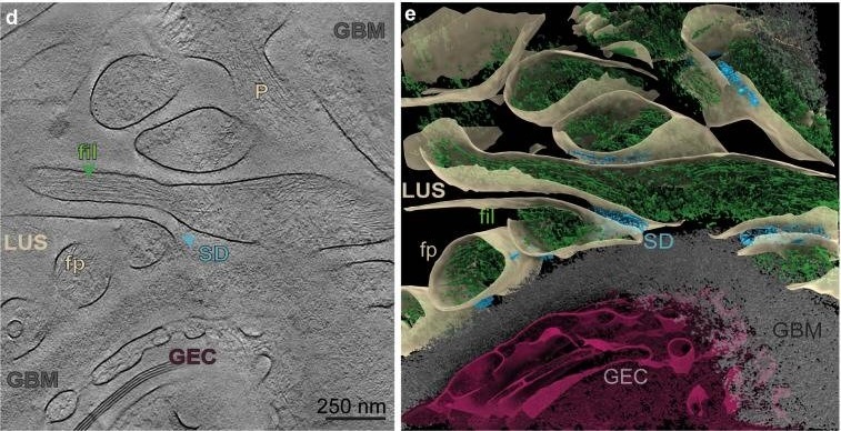
A comprehensive understanding of the molecular architecture of the slit diaphragm (SD), a critical component of the glomerular filtration barrier, is essential in elucidating renal filtration physiology and the mechanisms underlying kidney disease. Disruption of the SD is a hallmark of all forms of glomerulopathy, regardless of whether the cause is genetic, immunological, metabolic, or vascular.Our recent work demonstrated that the SD architecture in mice and Drosophila resembles a fishnet, with species-specific structural adaptations. Using cryo-electron tomography of FIB-milled samples, we resolved the bilayered fishnet architecture of the SD and showed that the main structural proteins Sns and Kirre (the Drosophila orthologs of nephrin and Neph1) organise into criss-crossing strands spanning the extracellular space between adjacent plasma membranes.
Figure 1. Cryo-electron tomography of the Drosophila nephrocyte slit diaphragm.
(a) Fluorescence microscopy. (b) FIB-SEM lamella. (c) Colour-annotated cryo-ET slice. (d) High-magnification cryo-ET slice. (e) 3D segmentation. From Moser, Lang, Birtasu et al., Nature Communications 2025.
Figure 2. Molecular architecture of the slit diaphragm.
MD simulation of Nephrin-Neph1 Ig domain complexes and idealized molecular model with interaction interfaces. From Birtasu et al., Kidney International 2025.
Mission of the PhD Candidate
The candidate will analyse disease models of minimal change disease and FSGS using nephrocytes from Drosophila melanogaster or zebrafish, combining cryo-FIB-milling and cryo-ET with quantitative structural analysis.
- Prepare serial cryo-lamellae from disease models of minimal change disease and FSGS by cryo FIB-milling
- Analyse disease progression by cryo-electron tomography and correlative light and electron microscopy (CLEM)
- Determine structural differences in slit diaphragm architecture by subtomogram averaging
- Develop algorithms for automated segmentation and quantitative analysis of the SD architecture
- Correlate structural findings with functional filtration data to establish structure-function relationships
Candidate Profile
Required
- Excellent MSc in molecular biology, biochemistry, structural biology, biophysics, or related discipline
- Strong interest in electron and/or light microscopy
- Experience with cell biological or biochemical techniques
- Very good written and spoken English
- Motivation for interdisciplinary work
- Hands-on experience with cryo-EM, cryo-ET, or FIB-milling
- Familiarity with RELION, IMOD, or ChimeraX
- Programming skills (Python, MATLAB)
- Experience with Drosophila or zebrafish models
- Interest in structural biology and computational methods
What We Offer
- The position is embedded in the DFG Research Training Group iMOL (GRK 2566), providing a structured PhD programme including lectures, workshops, retreats, and an annual Winter School.
- You will have access to state-of-the-art cryo-electron microscopes and a FIB-SEM at the Frankfurt Centers for Advanced Light Microscopy (FCAM) and Electron Microscopy (FCEM).
- The position is funded at E13 TV-G-U, 65 %, for 36 months.
Please send your application (cover letter, CV, transcripts, names of two referees) as a single PDF.
Indicate your interest in this specific project in your cover letter to:
email
Indicate your interest in this specific project in your cover letter to:
Bitte beziehe dich bei deiner Bewerbung auf jobvector
Für diesen Job einen passenden Lebenslauf erstellen und direkt bewerben
Lebenslauf erstellen
